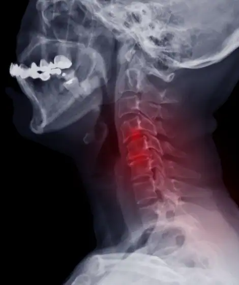
What is cervical spondylolisthesis?
Cervical spondylolisthesis is a common condition where a vertebra of the cervical spine is not properly aligned with its neighbor, allowing the bones to slip (or “translate”) with movement and causing pressure on the adjacent nerve roots and spinal cord. This can result in pain, numbness, weakness, gait problems, and bowel and bladder issues. In most cases, spondylolisthesis happens due to aging and long-term microtrauma. As the discs and ligaments of the spine age, they lose their elasticity and are unable to maintain normal alignment with physiologic movement.
What are the signs and symptoms of cervical spondylolisthesis?
Misalignment of the bones of the cervical spine can result in pressure on the adjacent nerves and spinal cord. Cervical spondylolisthesis can result in nerve root symptoms (radiculopathy) resulting in arm pain, weakness, numbness (“pins and needles sensation”), muscle spasm, and impaired coordination. If the spinal cord is compromised, then cervical myelopathy symptoms may result (gait difficulty, arm and leg weakness, pain, spasticity, impaired sensation, bowel and bladder difficulties, and impaired sexual function). Symptoms may be worse with neck movement if overt spinal instability is present.
How is cervical spondylolisthesis treated?
Mild symptoms associated with cervical spondylolisthesis can be treated with conservative measures (physical therapy, anti-inflammatory agents, oral steroidal agents, epidural steroid injections, dry-needling, etc.). If overt spinal cord compression is present or if conservative measures fail to provide adequate relief, anterior cervical decompression with fusion, posterior cervical decompression, or (in severe cases) combined anterior-posterior decompression, fusion, and instrumentation may be indicated.
What should you not do when you have cervical spondylolisthesis?
Patients with cervical spondylolisthesis should avoid high-impact activity affecting the neck, such as high-impact aerobics, riding ATVs and snowmobiles, skiing, etc.
What is cervical foraminal stenosis?
At every level in the cervical spine, openings between adjacent bones allow the passage of cervical nerves. These supply the neck and arms with sensation, motor function, and autonomic supply (including control of blood vessel tone and sweating). Herniation of a cervical disc or the formation of bone spur (osteophyte) can narrow the openings, thus compressing the nerves and causing cervical radiculopathy. This can result in pain, motor weakness, and/or loss of sensation in the distribution of the involved nerve. As cervical foraminal stenosis is due to osteoarthritis in most cases, it tends to progress with time.
How is cervical foraminal stenosis treated?
Treatment of cervical foraminal stenosis involves creating more room for the cervical nerve to pass through the foramen, either by shrinking inflammation within the nerve itself or physically expanding the foramen. Conservative measures include physical therapy, steroidal agents, non-steroidal anti-inflammatory agents, chiropractic care, traction, and epidural steroid injections/selective nerve root blocks. Patients who don’t improve after conservative management can opt for anterior or posterior cervical surgery. In our experience, surgery for symptomatic foraminal stenosis is effective in over 99% of patients with a complication rate of less than 1.5%.
Will cervical foraminal stenosis get worse?
As cervical foraminal stenosis is due to osteoarthritis in most cases, it tends to get worse with time as osteophyte formation progresses.
What is cervical radiculopathy?
Radiculopathy refers to disease or malfunctioning of a cervical nerve root, most commonly due to compression by arthritic spurs (osteophytes) or by a herniated disc. This results in irritation and malfunctioning of the nerve, causing pain in the nerve’s distribution (referred to as “radicular pain”); weakness in the muscle groups (the “myotome”) that the nerve supplies; loss of sensation, numbness, or paresthesias (“pins and needles sensations”) in the distribution (the “dermatome”) that that nerve supplies. Cervical radiculopathies can also occur as the result of trauma (fractures), compression by tumor or misplaced screws, viral infections, abnormal blood vessels, etc. In most cases, cervical radiculopathies improve with decompression of the affected nerves.
What is cervical myelopathy?
Cervical myelopathy refers to malfunctioning of the spinal cord, usually due to compression, most commonly caused by osteoarthritis or herniated cervical discs. It is more serious than cervical radiculopathy, as the spinal cord does not respond readily to decompression. Symptoms of spinal cord compression include ill-defined pain, weakness, loss of sensation, coordination difficulties, gait problems, impaired bowel and bladder function, and diminished sexual function. Symptoms can occur over a long period of time and not be recognized until the spinal cord compression is advanced. Signs of spinal cord compression include abnormally brisk reflexes, weakness, spasticity, impaired sensation, increased muscle tone, impaired ability to walk, spontaneous fasciculations of the muscles, and a whole host of pathological neurological reflexes.
What is cervical myelomalacia?
When the spinal cord is damaged by compression or impaired blood flow, an area of damage (scar tissue referred to as “gliosis”) can develop. The region of damage can be visualized on an MRI as a white spot. These may or may not be reversible with surgical decompression. Long term myelomalacia is usually associated with atrophy of the spinal cord and has a very poor prognosis for overall recovery. Myelomalacia is clinically associated with the development of a significant cervical myelopathy. At any rate, the appearance of myelomalacia on MRI suggests a significant concern for spinal cord dysfunction and should be fully evaluated to diagnose the exact cause to avoid further neurologic deterioration. Nonsurgical causes of myelomalacia include demyelination (loss of the insulation of the spinal cord nerve fibers) as seen in multiple sclerosis or cord changes associated with viral infections and autoimmune diseases.
What is cervical lordosis?
Normal lordosis of the cervical spine critically limits normal movement of the neck, preventing misalignment during physiologic movement. As people age, lordosis generally decreases, leading to chronic neck pain. Exaggeration of the normal cervical lordosis is referred to as “hyperlordosis” and is a form of scoliosis (“curvature of the spine”). Causes of hyperlordosis include trauma, poor posture, osteoporosis, obesity, cervical spondylolisthesis, neuromuscular disorders, and certain congenital disorders involving the bones and connective tissues. In certain cases, surgery may be required to correct normal cervical alignment to eliminate pain and decompress the spinal cord and nerves.
What is cervical kyphosis?
Cervical kyphosis refers to malalignment of the neck bones with reversal of the normal C-shaped curve from the skull base to the upper chest. In kyphosis, the spine curves backwards instead of forward. This can result in chronic pain due to muscle fatigue. Cervical kyphosis is common in osteoarthritis affecting the spine, and can also be seen after past surgery and trauma, various neuromuscular disorders; tumor involvement; and in cases of congenital bone and connective tissue disorders. Surgery may be advised in certain cases. Severe cases of kyphosis result in “chin on chest deformity.” Reversible kyphosis can also be seen with cervical muscle spasm due to a cervical radiculopathy. This can be corrected with appropriate treatment. In cases involving cervical surgery, correcting a kyphosis to reattain a normal “sagittal balance” is important for the cervical spine to function normally postoperatively with no pain.
What is OPLL (ossification of the posterior longitudinal ligament)?
Ossification of the posterior longitudinal ligament (OPLL) is a condition characterized by abnormal bone formation involving the ligaments along the posterior aspect of the cervical vertebral bodies within the spinal canal. It can lead to severe spinal cord compression and cervical myelopathy. Because the abnormal bone formation fuses the ligament and the dura (the membranous covering of the spinal cord) into a solid sheet of bone anterior to the spinal cord, surgical decompression may be exceedingly difficult. Surgery for OPLL has a significantly higher risk of spinal cord injury and cerebrospinal fluid leak post-operatively and usually requires a combined anterior-posterior decompression, fusion, and instrumentation.
What is cervical instability?
Spinal instability refers to hypermobility of a spinal segment (“toggling” of the bones) resulting in progressive spinal deformity and/or spinal cord and nerve root compression and associated neurologic defects. In the cervical spine, this can result in cervical radiculopathy, myelopathy, or myeloradiculopathy. In technical terms, a segment that shows movement over 3.5mm or angulation of over 11 degrees on flexion-extension X-rays is considered unstable. Overt instability or progressive neurologic deficits require surgical decompression and fusion. Causes of cervical instability include trauma; previous surgery; degenerative osteoarthritis; degenerative joint conditions like rheumatoid arthritis; various neuromuscular disorders; connective tissue disorders; involvement of the bones due to tumor, etc.
What is cervical swan neck deformity?
In severe cervical kyphosis cases, an “S” shaped deformity of the cervical spine (resembling the neck of a swan) can result. As this deformity progresses, spinal cord and nerve root compression can occur, resulting in neurologic deficits. The most common cause of a swan neck deformity is severe cervical osteoarthritis as well as post-traumatic and after prior cervical surgery (especially after a cervical laminectomy without fusion and instrumentation). Surgical indications include progressive deformity, intractable pain, and neurologic deficits. Surgery requires decompression of the spinal cord and nerves, and recreation of normal sagittal balance with reconstruction of the normal cervical curvature (lordosis). In many cases, this requires a multilevel cervical corpectomy with instrumentation and fusion anteriorly and a cervical fusion and instrumentation posteriorly. Surgery for a swan neck deformity is complex and is best performed by an experienced cervical spine surgeon.
How is cervical instability diagnosed?
Diagnosis of cervical instability requires radiographic demonstration of too much movement (translational instability) or too much angulation (angular instability) at a particular spinal segment. Historically this is done using cervical spine X-rays with the patient’s neck in flexion and extension positions (flexion-extension X-rays). In addition, MRI of the cervical spine with the patient’s neck in both flexion and extension is especially useful as they can visualize actual compression of the spinal cord and nerves, changes in bony alignment, and changes in disc bulging and ligamentous compression with the dynamic movements of the patient’s neck.
What is cervical pseudoarthrosis?
Pseudoarthrosis is a failure of the bones to fuse after attempted fusion surgery. This can lead to abnormal motion between the bones, resulting in chronic pain in some patients. It can progress to the point of instability, resulting in recurrent nerve root and spinal cord compression. Any factor that reduces the ability of bone-forming cells (osteoblasts) to form new bone can increase the risk of pseudoarthrosis. The most common cause of pseudoarthrosis after attempted cervical surgery is smoking or continued use of tobacco products. Other factors include malnutrition, obesity, advanced age, osteoporosis and other bone disorders, diabetes, rheumatoid arthritis, and use of steroids and non-steroidal anti-inflammatory agents. Patients who undergo multilevel fusion surgery or who are too active after their fusion surgery also have a higher risk of pseudoarthrosis. Diagnosis is made with flexion-extension films. If conservative measures fail to alleviate pain, repeat surgery may be indicated.